
Our feet and ankles are pivotal in keeping the rest of the body stable and mobile, as they absorb the impact of walking, running and jumping, and ensure that we can stand without putting unnecessary pressure on other muscles or joints.
The feet contain almost a quarter of the body’s bones, as well as hundreds of connective ligaments and tendons.
Combined with the complexity of the ankle joint, both are very susceptible to injury, with the ankle being the most commonly injured joint and a physiotherapist most common injury area.
We want to help keep you on your feet with our extensive knowledge and treatment options for feet and ankle pain. Like with any other part of the body, we should listen to our feet and notice the changes we may feel.
Are my feet feeling more fatigued? More sore? More swollen? Regardless of the issue, our Pivotal Motion Physiotherapy team will be able to help you!

PRIMARY FUNCTION OF OUR LOWEST WEIGHT BEARING JOINTS
The main function of the feet and ankle is to provide stability as we move around, whether on the sporting field or during activities of daily living. The feet and ankles assist in supporting the body’s weight, ensuring other joints such as the hips and knees are able to function properly.
The feet and ankles are also one of the most vital components of posture and balance, as their stabilisation assist the rest of the body. Through the anatomy of the foot, the sole of the foot enables other joints to keep stable without overextending.
ANATOMY, BIOMECHANICS, INJURIES & SYMPTOMS OF THE FOOT AND ANKLE
As we are constantly on our feet, ankle and foot injuries can be commonplace, especially when participating in sports and physical activity.
ANKLE JOINT
Swelling and pain when moving are key symptoms of a foot or ankle injury, as is not being able to support any weight. Stress fractures, often a result of over training, can also be common, and can worsen if not properly rested.
ANATOMY OF THE FOOT AND ANKLE
The two, key movements available at the ankle are dorsi- and plantar flexion (pointing foot up or down), and inversion and eversion (side-to-side). These movements are dictated by the structures of the joints making up the ankle complex. The joints allow these movements are the talocrural joint and the subtalar joint.
The talocrural joint is made up of the articulations between the tibia (shin bone), fibular (outer bone of the lower leg) and the talus (upper bone of the foot). This joint is called a synovial hinge, and allows the dorsi- and plantarflexion at the ankle.
The second joint of concern is the subtalar. The articulation between the talus and our heel bone, the calcaneus, this joint provides the side-to-side movements of the foot which allow our smooth transitions over different terrains.
These joints are provided some structural stability by their bony formation, however get further passive support from a number of ligaments, which work to prevent excessive translation of the bones in any direction. Due to the importance of movement at the ankle, there are also a lot of muscular attachments around the area, hence tendon injuries can also affect the joint function and stability.
BIOMECHANICS OF THE FOOT AND ANKLE
Alignment of the feet and ankle have significant ramifications for not only foot and ankle health but, reducing the risk of knee, hip and spinal complications. It can affect posture as well as our functional movements such as walking and running. Pes planus or ‘flat feet’, refers to the arch of the foot being reduced. Pes planus is not only a visual ailment of the foot but means that the rest of the body has an unstable base, which alters the alignment through the knee and hip.
This misalignment changes where the nature of forces are placed through our joints, which can lead to many complications throughout the lower limb and back. Physiotherapist are professionals in identifying abnormal movement patterns and providing strategies to correct any insufficiencies.

INJURIES
FRACTURES OF THE ANKLE
Ankle fractures are one of the less common ankle injuries and can occur at the distal tibia or fibular, talus or calcaneus. Ankles fractures will normally occur when a large, twisting, multi-directional force is applied to the ankle. A fractured ankle may present similarly to a severe ligament sprain, the onset of pain will be immediate and intense, with swelling, bruising and inflammation occurring soon after. The ankle will usually be unable to bear weight and range of motion will be limited, there may also be joint deformity if the fracture is severe enough.
DISLOCATIONS OF THE ANKLE AND FOOT
A dislocated ankle is not an injury which is commonly seen and it is usually partnered with a fracture. Due to the structure of the joint and the positioning of the mortise made by ends of the tibia and fibular bones (medial and lateral malleolus respectively), a full dislocation will rarely occur independent of fracture.
Dislocation will generally occur due to severe trauma, possible mechanisms include a serious sprain, sporting injury, fall or motor vehicle accident. Symptoms of a dislocation will include intense pain, deformation of the joint, swelling, bruising and inflammation, an inability to bear weight and a reduction in movement.
It is very important that you present to a medical practitioner immediately if an ankle dislocation is suspected. As there are a range of neurovascular structures positioned around the area and the amount of damage that occurs with a dislocation, a large concern is that blood vessels or nerves will be damaged with the injury.
Present to your relevant health professional and they will be able to assess the injury and aid in recovery. Physiotherapy will be important in full recovery from a dislocated ankle as the joint will need to be reloaded and there will be a large amount of muscular and ligament damage to remedy as well. Our physio’s will help to return maximal functionality back to the joint and help you to reach realistic and timely recovery goals throughout the process.
LIGAMENTS INJURY
Ligament damage is the most common of ankle and joint injuries. Damage to the ankle ligaments is what is known as a sprained ankle and can be graded from grade 1-3 depending on the severity of the injury. The ankle has several ligaments that can be injured depending on the type of injury occurred.
The most common ankle sprain is a lateral ankle sprain, by where, the ligaments on the outside aspect of the ankle are injured when the ankle has inverted or “rolled” inwards and plantarflexed. The most injured ligament is the anterior talofibular ligament (ATFL) which connects the talus and fibular on the anterior-lateral aspect of the ankle. The symptoms of an ankle sprain include history of an inversion plantarflexion injury, pain, swelling, bruising and instability.
Physiotherapy will work to initially reduce inflammation through the ankle before restoring range of motion and muscle function. A key component to recovering from an ankle sprain includes regaining balance and proprioception. Your physiotherapist will be able to give you a specialised exercise program, as well as, apply manual therapy techniques to maximise your recovery.
ACHILLE’S TENDON
ACHILLE’S TENDINOPATHY
The Achilles tendon is the largest tendon in the body which originates from the gastrocnemius and soleus muscles (calf muscles) and attaches to the back of the calcaneus (heel).
Achilles tendinopathy is a common overuse injury that occurs due to repetitive microtrauma of the tendon. Without adequate healing time the microtrauma progresses causing significant degeneration of the tendon, tendon sheath or both.The type of load is key to why Achilles tendinopathy can present and excessive pronation at the foot has been heavily linked with tendinopathy. Early intervention and load management will ensure the tendons health remains optimum to avoid aggravation.
ACHILLE’S RUPTURE
The achilles tendon is the most commonly ruptured tendon in the human body and is a common sport injury. It primarily occurs during stop-go motions in recreational sporting activities. There is often an audible sound and reports of feeling a sharp snapping pain at the back of the ankle. The risk of an acute rupture of the achilles tendon often arises from a progressive degeneration of the tendon and can be because of chronic achilles tendinopathy. An achilles rupture is usually managed surgically and early intervention, physiotherapy is also vital in returning to pre-morbid function.

FEET
The foot can be broken into 3 separate regions to help simplify the area of concern; rear foot, mid foot and forefoot (picture of bony anatomy). The foot is a flexible structure made up of bones, ligaments and soft tissue which allow it to provide us with enough mobility, as well as, stability to perform tasks such as running, walking, jumping etc.
REAR FOOT
The rear foot is comprised of the calcaneus (heel) and talus (ankle). The talus provides the support for the lower leg bones and forms the lower half of the ankle joint. The most common cause of rear foot (heel) pain is plantar fasciitis. Other causes include stress fractures of the calcaneus (heel), fat pad syndrome and tarsal tunnel syndrome.
PLANTAR FASCIITIS
Plantar fasciitis physiotherapy is used to treat the condition where the plantar fascia is compromised, usually resulting in medial heel pain. The plantar fascia is a strong band of tissue that runs through the bottom of your foot from your heel bone (the calcaneus) to the joints in the ball of your foot. Its primary function is to support the bottom arch of the foot, whilst providing shock absorption with its elastic properties.
Plantar fasciitis often presents as a chronic condition that has developed over time, leading to significant degenerative changes to the soft tissue. It presents with increased pain immediately post periods of rest (getting up in the morning or after sitting for prolonged time). A typical presentation is seen when the initial morning pain slowly recedes throughout the day, without ever completely going away.
Physiotherapy has been shown to be effective in decreasing pain in patients with plantar fasciitis. The techniques used include; soft tissue release, joint mobilisation, taping, ice and a staged rehabilitation exercise program. Physiotherapy can get those with plantar fasciitis back to function quick with a reduced chance of re-aggravation.
MID FOOT
The mid foot is includes a series of bones (3 cuneiforms, navicular, cuboid) as well as soft tissue, that help provide the arch within our foot. When a load is placed through the foot the bones are compressed together and provide a strong dynamic support structure for the foot. When the load is removed, the bones distract away, allowing movement and flexibility in the foot. The most common cause of pain in the mid foot is mid tarsal joint pain following an ankle injury. Other common causes of mid foot pain include stress fractures of the navicular, usually due to an overuse stress injury.
NAVICULAR STRESS FRACTURE
The navicular is a bone in the mid foot which is on the medial side of the foot. It is one of the most common stress fractures seen in the foot – specifically in the exercising population. Stress fractures are commonly caused due to repetitive overloading without an adequate time for healing to occur. A look into movement patterns and biomechanics during repetitive activities give insight into potential causes and room for change to avoid further injuries. The pain is often vague and hard for someone to pin point themselves. The pain can radiate along the medical arch and into the top of the foot.
Following a navicular stress fracture rest is important to allow the bone to heal. The rest is usually with some weight bearing in a cast for 6-8 weeks. After this period of rest, regaining movement and strength around the ankle is vital for full recovery. Decreased range in the ankle has been shown to be a risk factor for developing a navicular stress fracture as compensatory movements occur in the mid foot to achieve full function. Physiotherapy works to correct any predisposing factors that can lead to a stress fracture and developing a gradual return to function program.
LISFRANC INJURY
One of the most significant injuries that can occur in the mid foot region is a Lisfranc injury which involves a fracture or dislocation at the first tarsometatarsal joint (1st cuneiform and 2nd metatarsal base). The mechanism of injury usually involves a compressive force on the back of the heel forcing the toes to be forced backward whilst the foot is trapped.
Following a Lisfranc joint injury pain is often present in the mid foot with difficulty accepting weight onto the injured foot. Specifically force to the forefoot can be aggravative (pushing off toes whilst walking, standing on toes, calf raises). Lisfranc injuries can include partial strains, complete ruptures, fractures and dislocations depending on the severity.
Depending on the severity and instability present within the injured joint management of the area will be altered. Early detection is key and plain x-rays during weight bearing are recommended to identify the severity. In mild cases conservative management with non-weight bearing for an initial period of 6 weeks is recommended to allow time for tissue healing.
Following this period of immobilisation, strengthening and mobilising of the ankle is necessary to achieve full function. In more severe cases review from an orthopaedic specialist is recommended and surgical fixation may be required to stabilise the joint. Your physiotherapist will work closely with the surgical team to help devise a graded return to function plan and achieve your rehabilitation goals.
FOREFOOT
The forefoot includes the five toes (phalanges) and five long bones (metatarsals) in the foot. Many complications with the forefoot involve pressure areas creating corns, calluses and nail complications. The forefoot includes one of the most important joints in the foot, the first metatarsal phalangeal joint (first toe). This joint is responsible for a lot of function during walking, running and must adhere to large amounts of force in these activities. With limited range of movement in the first MTP a normal pattern of movement is unable to be achieved, resulting in altered biomechanics which can lead to additional complications.
HALLUX LIMITUS/RIGIDUS
Hallux limitus refers to a restriction in dorsiflexion of the hallux (big toe) at the MTP joint. This limitation in movement is primarily due to degenerative arthritic changes within the joint. Hallus rigidus is a term used for the final progression of this condition by which the joint is relatively immobile and some fusion of the bones has occurred.
The primary role of the hallux is to allow dorsiflexion during walking/running. When this range is limited the foot is forced to adapt and several compensatory movements can occur to help achieve the desired result. Pain is usually present right in the MTP joint and is aggravated during walking, as well as, any forefoot weight bearing.
Several biomechanical changes can occur along the lower limb chain, at the knee and hip, which can result in secondary injuries. Treatment of this condition begins by reducing the pain and force at the painful joint. From there addressing any biomechanical issues is important. A full biomechanical analysis and footwear assessment can help identify strategies to decrease the chance of condition progression and allow full function.
HALLUX VALGUS
Hallux valgus, commonly known as ‘bunions’, is a progressive foot irregularity whereby, there is deviation of the first toe at the metatarsophalangeal (MTP) joint. The lateral deviation of the big toe is a sign of the gradual subluxation of the MTP joint and is usually first noticed due to the presentation of the foot deformity. In the early stages of hallux valgus there is often no symptoms, but as the condition progresses, pain is usually felt on the medial aspect of the bunion. The use of restrictive footwear (high heels) and excessive pronation (flat feet) has been shown to both increase the risk of developing the condition, as well as, progress symptoms once first identified.
Treatment for hallux valgus is focused on correcting alignment and de-loading the injured area. By correcting possible causative factors the condition can be managed well conservatively. In severe cases surgical techniques can be completed, but, there is little evidence to support the use of them.
INJURY AVOIDANCE & RECOVERY FOR THE FEET AND ANKLE
 As we spend so much time on our feet, an injury to the ankle or the foot itself can severely limit our day-to-day lifestyle, in addition to our fitness.
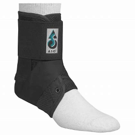
Rest and keeping weight off the foot is the best way to help recovery. Strapping tape or other forms of ankle support can assist in reducing the likelihood of reinjuring, as the added stability ensures that the ligaments and tendons aren’t overextended.
The correct fitting shoe can also ensure a reduced risk of injury, as most running shoes add support to both the ankle and sole of the foot. Combined with regularly physiotherapy and podiatry appointments, the ankle and foot can be strengthened. Podiatry insoles can also reduce the negative effects of poor foot arches, improving the support and stability of the rest of the body.
If you are suffering ankle or foot pain it is important to get treatment as soon as possible to maximise recovery!
Call us now on 07 3352 5116 or book online to see one of our experienced physiotherapists.